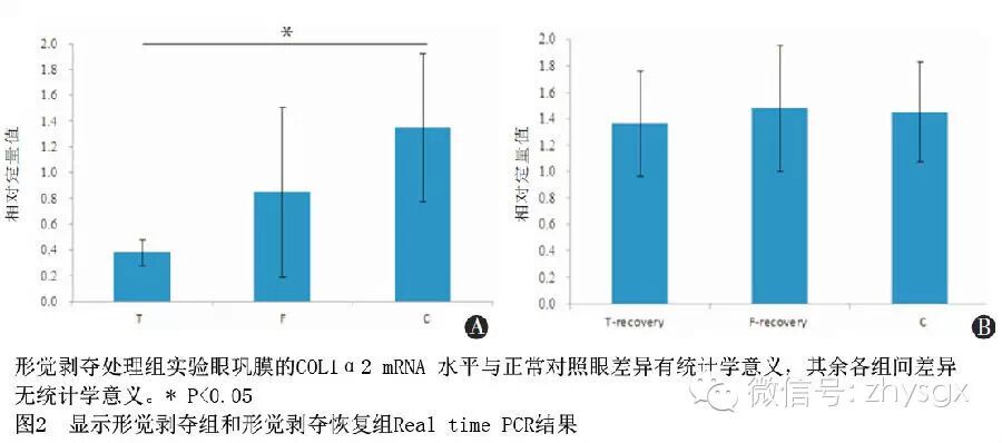
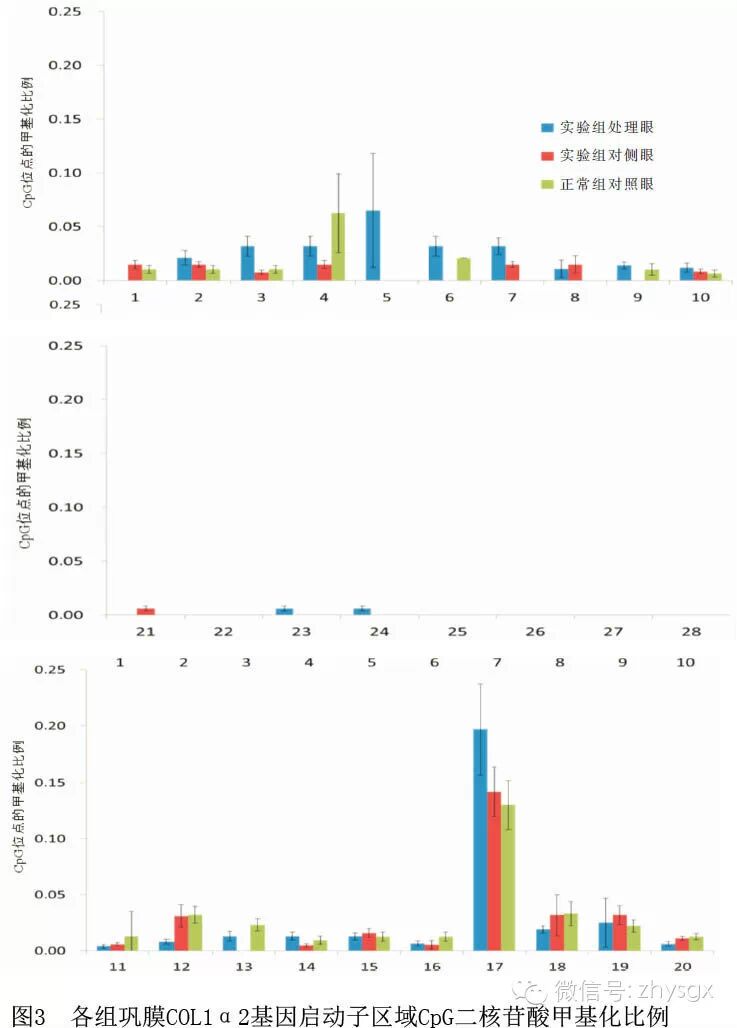
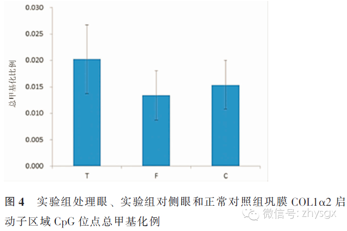
【摘要】 目的 研究C57BL/6小鼠形觉剥夺性近视形成和恢复期巩膜COL1α2 mRNA表达及其启动子区域CpG岛甲基化水平。方法 实验研究。单眼形觉剥夺建立C57BL/6小鼠近视动物模型和恢复期动物模型,分别单眼遮盖4周(48只)和单眼遮盖4周恢复1周(24只),另外设立正常对照组小鼠36只。实验前后分别用红外偏心摄影验光仪测量小鼠眼球的屈光状态,OCT检测小鼠眼轴长度和玻璃体腔深度。RT-PCR检测巩膜COL1α2 mRNA表达水平;硫化测序聚合酶链式反应(BSP)检测C57BL/6小鼠巩膜COL1α2启动子区域CpG岛甲基化水平。同一小鼠的实验眼和对侧眼比较采用配对t检验,不同组间比较采用独立样本t检验。结果 形觉剥夺4周,实验眼相比对侧眼形成明显的相对近视(t=-2.64,P<0.05),并伴有眼轴和玻璃体腔延长。形觉剥夺4周恢复1周后,相对近视和延长的眼轴和玻璃体腔长度都获得恢复。形觉剥夺4周后,实验眼和正常对照眼相比COL1α2 mRNA表达明显下降(t=3.05,P<0.05),形觉剥夺4周恢复1周实验眼与正常对照眼相比差异无统计学意义。形觉剥夺4周,实验眼和对侧眼及正常对照眼COL1α2启动子区域CpG岛甲基化水平差异均无统计学意义。结论 小鼠单眼形觉剥夺4周可诱导出相对近视,巩膜内COL1α2 mRNA表达明显下降,但该改变与其基因启动子区CpG岛的甲基化状态无关。
【关键词】 近视; COL1α2; 启动子; DNA甲基化; 模型,动物
DOI:10.3760/cma.j.issn.1674-845X.2014.06.002
基金项目:国家973计划(2011CB504604);国家自然科学基金(81070751、81170870、81271039);教育部新世纪人才项目(NCET-10 0977);中组部青年拔尖人才支持计划
作者单位:325027
通信作者:周翔天,Email:zxt-dr@wz.zj.cn
Study of CpG methylation of the COL1α2 promoter in form-deprivation myopia in C57BL/6 mice Jiao Shiming, Zheng Fan, Ying Huangfang, Huang Furong, Zhao Fuxing, Qu Jia, Zhou Xiangtian. Eye Hospital of Wenzhou Medical University, Wenzhou 325027, China
Corresponding author:Zhou Xiangtian,Email:zxt-dr@wz.zj.cn
【Abstract】 Objective To investigate the changes in refraction and ocular biometric parameters in form-deprivation myopia; to try to find the changes in mRNA and methylation in the promoter of the COL1α2 gene during the form deprivation and recovery periods. Methods This was an experimental study to establish the C57BL/6 mouse animal model for form-deprivation myopia and recovery. Mice in the form-deprivation group wore a diffuser on a randomly chosen eye for 4 weeks (n=48) and another group underwent 4 weeks of monocular diffuser treatment followed by 7 days of recovery (n=24). An additional 36 mice without any treatment were the normal control group. Refraction was measured by photoretinoscopy. Vitreous chamber depth (VCD) and axial length (AL) were measured by optical coherence tomography (OCT) with focal plane advancement. The COL1α2 mRNA level in the sclera was determined by real-time PCR and the COL1α2 methylation state was observed by bisulfite DNA sequencing. Data were analyzed using t test. Results After 4 weeks of form deprivation, a significant myopic shift had been induced in the treated eyes compared with the fellow eyes. Before and after the experiment, refractions were -1.89±1.97 D and -3.18±1.09 D (P<0.05). After 28 days of monocular deprivation (MD), the promoter region of COL1α2 was methylated more frequently in the deprived eyes than in the controls. However, there were no significant differences among the form deprived, fellow and normal control eyes. Conclusion Form-deprivation myopia can be induced in C57BL/6 mice. Scleral COL1α2 mRNA was reduced in MD eyes compared with normal controls, but this change and its gene promoter-region methylation status of CpG island are not related.
【Key words】 Myopia; COL1α2; Promoter; DNA methylation; Models,animal