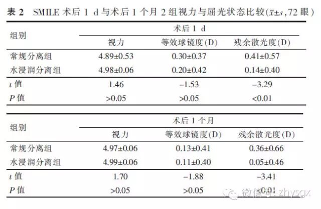
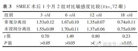
【摘要】 目的 观察水浸润分离法在SMILE术中的安全性和有效性。方法 前瞻性临床对照研究。将拟采用SMILE手术完成近视及近视散光患者72例(144眼)纳入研究,每例患者随机选取一眼常规分离角膜基质透镜,对侧眼水浸润分离角膜基质透镜。观察术中切口上皮损伤情况,光镜下观察透镜边缘的整齐程度。采用独立样本t检验比较2组术后视力、屈光状态、对比敏感度。结果 术中发生角膜切口边缘上皮损伤水浸润分离组与常规分离组分别为2眼(3%)与14眼(19%),差异有统计学意义(χ2=6.41,P<0.01)。采用水浸润法分离的角膜基质透镜边缘光滑整齐,常规分离的透镜边缘相对粗糙。水浸润法分离组与常规分离组术后第1天视力为4.98±0.06与4.89±0.53,等效球镜度为(+0.20±0.42)D与(+0.30±0.37)D,差异无统计学意义(t=-1.53,P>0.05),残余散光度为(0.14±0.40)D与(0.41±0.57)D,差异有统计学意义(t=-3.29,P<0.05)。术后第1个月视力分别为4.99±0.06与4.97±0.06,等效球镜度分别为(+0.11±0.40)D与(+0.13±0.41)D,差异无统计学意义(t=-1.88,P>0.05),残余散光度为(0.05±0.46)D与(0.36±0.66)D,差异有统计学意义(t=-3.41,P<0.05)。2组对比敏感度差异无统计学意义。结论 与常规SMILE术中微透镜分离法相比,水浸润分离法可有效地减少术中角膜上皮的损伤,减小分离阻力,取出的角膜基质透镜表面更加平滑,边缘更加整齐,并明显减少了术后残余散光。
【关键词】 飞秒激光; 飞秒激光小切口基质透镜取出术; 水浸润; 透镜分离
DOI:10.3760/cma.j.issn.1674-845X.2014.07.007
作者单位:400042 重庆,第三军医大学大坪医院野战外科研究所眼科
通信作者:白继,Email:baiji_liujing@163.com
Liquid infiltration method to separate lenticule in small incision lenticule extraction
Zhang Guowei, Chen Kaijian, Bai Ji, Kan Qiuxia, Liu Lina, Xu Duo, Lang Min. Department of Ophthalmology, Daping Hospital, Research Institute of Surgery, the Third Military Medical University, Chongqing 400042, China
Corresponding author:Bai Ji,Email:baiji_liujing@163.com
【Abstract】 Objective To observe the safety and efficacy of liquid infiltration method to separate lenticule in small incision lenticule extraction (SMILE). Methods In this prospective study, 72 patients were included. Randomly selected one eye was performed conventional method; the contralateral eye was performed liquid infiltration method. The corneal epithelium injury around the incision in surgery and the smoothness of the lenticule edge was observed under optical microscope. The differences of visual acuity, refraction, contrast sensitivity postoperatively between the two groups using independent samples t test. Results The corneal epithelium injuries were observed in 2 eyes in liquid infiltration method and 14 eyes in conventional method during surgery, the difference was statistically significant (χ2=6.41, P<0.01), the lenticule edges of liquid infiltration method were smoother than conventional method. The visual acuity, spherical equivalent (SE) and residual astigmatism of liquid infiltration method and conventional method 1 day postoperative were: 4.98±0.06 and 4.89±0.53, +0.20±0.42 D and +0.30±0.37 D, 0.14±0.40 D and 0.41±0.57 D respectively. The difference of residual astigmatism was statistically significant(t=-3.29, P<0.05). The visual acuity, spherical equivalent and residual astigmatism of liquid infiltration method and conventional method 1 month postoperative were: 4.99±0.06 and 4.97±0.06, +0.11±0.40 D and +0.13±0.41 D, 0.05±0.46 D and +0.36±0.66 D, respectively. The difference of residual astigmatism was statistically significant (t=-3.41, P<0.05). The difference of contrast sensitivity was without statistically significant. Conclusion The liquid infiltration method makes the edge of lenticule smoother and effectively reduces the corneal epithelial injury and residual astigmatism. It is a safe and feasible skill in SMILE.
【Key words】 Femtosecond laser; Small incision lenticule extraction; Liquid infiltration; Lenticule separation