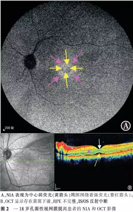
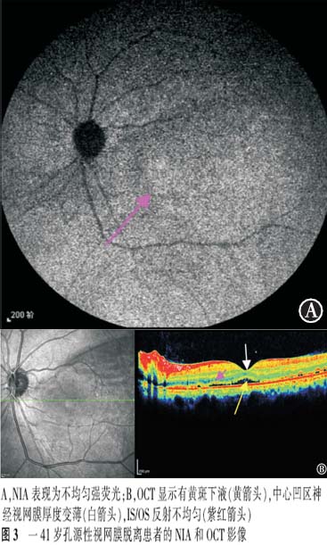
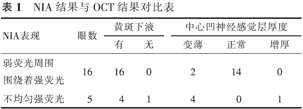
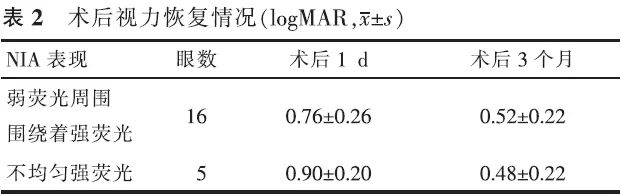
【摘要】 目的 近红外眼底自发荧光检查(NIA)联合OCT对孔源性视网膜脱离巩膜扣带术后早期黄斑结构的评估价值。方法 前瞻性系列病例研究。对21例(21眼)伴黄斑脱离的孔源性视网膜脱离患者行巩膜扣带术,于术后3个月行NIA及OCT检查。观察检查图像的异常和手术后视力的恢复情况。对视力的变化进行配对t检验,NIA与术后BCVA的相关性采用双向无序分类变量资料的关联性进行分析。结果 NIA:16眼表现为弱荧光周围围绕着强荧光,5眼表现为不均匀的强荧光。OCT:20眼存在黄斑下液;中心凹视网膜神经纤维层的厚度6眼变薄,14眼正常,1眼增厚;21眼IS/OS反射异常。视力恢复情况:术后3个月与术后1 d比较,NIA表现为弱荧光周围围绕着强荧光者,视力差异有统计学意义(t=9.922,P<0.05),NIA表现为不均匀强荧光者,视力差异无统计学意义(t=2.409,P>0.05),黄斑NIA表现与术后BCVA变化高度相关(C=0.502,P<0.05)。结论 孔源性视网膜脱离巩膜扣带术后早期,黄斑结构与功能均尚未完全恢复,需要我们加以重视。
【关键词】 视网膜脱离; 荧光; 荧光素血管造影术; 体层摄影术,光学相干; 巩膜扣带术
DOI:10.3760/cma.j.issn.1674-845X.2014.04.011
作者单位:030002 太原,山西省眼科医院眼科
通信作者:贾亚丁,Email:yadingjia@163.com
Melanin-related near-infrared fundus autofluorescence combined with optical coherence tomography for studying macular structure changes in the early stages after undergoing scleral buckling for rhegmatogenous retinal detachment
Ren Jingqin, Yuan Zhigang, Zhou Guohong, Hou Jia, Yang Jihong, Xie Juan, Xie Lisha, Chang Xin, Jia Yading. Shanxi Eye Hospital, Taiyuan 030002, China
Corresponding author:Jia Yading,Email:yadingjia@163.com
【Abstract】 Objective To evaluate the changes in macular structure in the early stages after undergoing scleral buckling surgery for rhegmatogenous retinal detachment (RRD); to use melanin-related near-infrared fundus autofluorescence (NIA) combined with optical coherence tomography (OCT) for studing the changes. Methods This was a prospective case-series study. Melanin-related near-infrared fundus autofluorescence (NIA) and optical coherence tomography (OCT) examinations were performed on 21 patients with local primary RRD three months after undergoing a successful scleral buckling procedure for retinal reattachment. The RRDs in all 21 eyes involved the macula. To check the anomalies fo the images and the recovery of the vision after surgery. Changes invision after surgery were compared with a paired t test. The correlation of NIA and BCVA was analyzed with the relevance of two-way disorderly classification variable data. Results NIA revealed that 16 eyes had hypofluorescence surrounded by hyperfluorescence and 5 eyes had uneven hyperfluorescence. Submacular fluid was found in 20 eyes. The retinal nerve fiber layer in the central fovea became thinner in 6 eyes. In one eye, the retinal nerve fiber layer in the central fovea became thicker, and in another 14 eyes the thickness of the retinal nerve fiber layer was normal. The reflection from the photoreceptor inner and outer segment junction (IS/OS) was abnormal in all 21 eyes. Visual acuity measured with NIA revealed that the hypofluorescence which was surrounded by hyperfluorescence had improved at the 3-month follow-up compared to the 1-day follow-up examination (t=9.922, P<0.05), while VA measured at the same time revealed that uneven hyperfluorescence had not improved at the 3-month follow-up (t=2.409, P>0.05). The macular NIA results and the change in postoperative BCVA were highly correlated (r=0.502, P<0.05). Conclusion Macular function and structure do not fully recover in the early stages after undergoing scleral buckling to treat RRD. More attention should be paid to this problem.
【Key words】 Retinal detachment; Fluorescence; Fluorescein angiography; Tomography,optical coherence; Scleral buckling